
Fingertip crush injuries are a devastating occurrence in both children and adults which occur during activities of daily living. At Dr. Amrika’s Aesthetics on Sarjapura Main Road, the focus is on quick and reliable restoration of function, with minimal pain and expenditure.
Mastering Fingertip Injuries: A Comprehensive Guide to Reconstruction, Flap Surgeries, and Salvaging Function
Section 1: Introduction to Fingertip Injuries
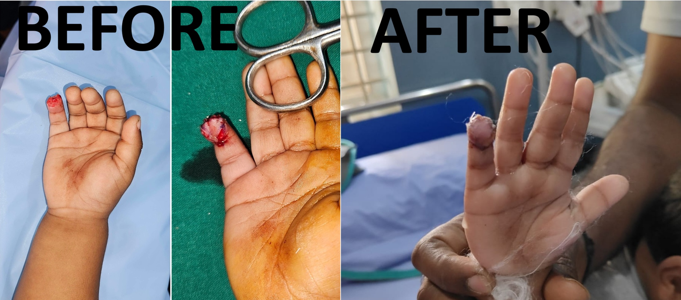
The human hand is a masterpiece of evolutionary engineering, and its most critical interface with the physical world is the fingertip. Far from being a simple cap at the end of a digit, the fingertip is a highly specialized organ of sensation, touch, exploration, and motor manipulation. It possesses a dense concentration of sensory receptors (including Pacinian and Meissner's corpuscles), a complex vascular network, a protective nail unit that provides counter-pressure to the pulp during pinch, and a vital structural scaffold provided by the distal phalanx.
Because of their constant involvement in daily activities, work, and exploration, fingertips are the most frequently injured parts of the upper extremity. They represent approximately two-thirds of all hand injuries in children and are a primary source of occupational disability in adults. Despite their frequency, these injuries are highly complex and demand a meticulous approach to management.
At our Level 3A Superspeciality center located on Sarjapura Main Road, Bengaluru, we approach every fingertip injury with a singular goal: to restore a stable, painless, and highly sensate digital tip while maintaining maximum length and aesthetic harmony. Led by Dr. Amrika Seshadri, a dedicated Plastic Surgeon with 11 years of standing, our clinic specializes in advanced soft-tissue reconstruction techniques.
It is important to clarify our scope of service from the outset: Dr. Amrika Seshadri does not perform microvascular replantation procedures. Instead, our center focuses entirely on the art and science of reconstructive salvage, utilizing local and regional tissue advancement flaps to repair, protect, and restore injured digits without the prolonged hospital stays, high donor-site morbidity, or systemic risks associated with microvascular digit reattachment.
This comprehensive guide serves as an extensive resource for patients, emergency physicians, and primary care providers to understand how fingertip injuries are classified, evaluated, and treated using state-of-the-art reconstructive techniques.
Section 2: Anatomy of the Fingertip
To understand the severity of a fingertip injury and the logic behind reconstructive options, one must first understand its intricate anatomy. The fingertip is defined anatomically as the portion of the digit distal to the insertion of the flexor digitorum superficialis (FDS) and extensor digitorum communis (EDC) tendons—essentially, everything from the distal interphalangeal (DIP) joint to the tip.
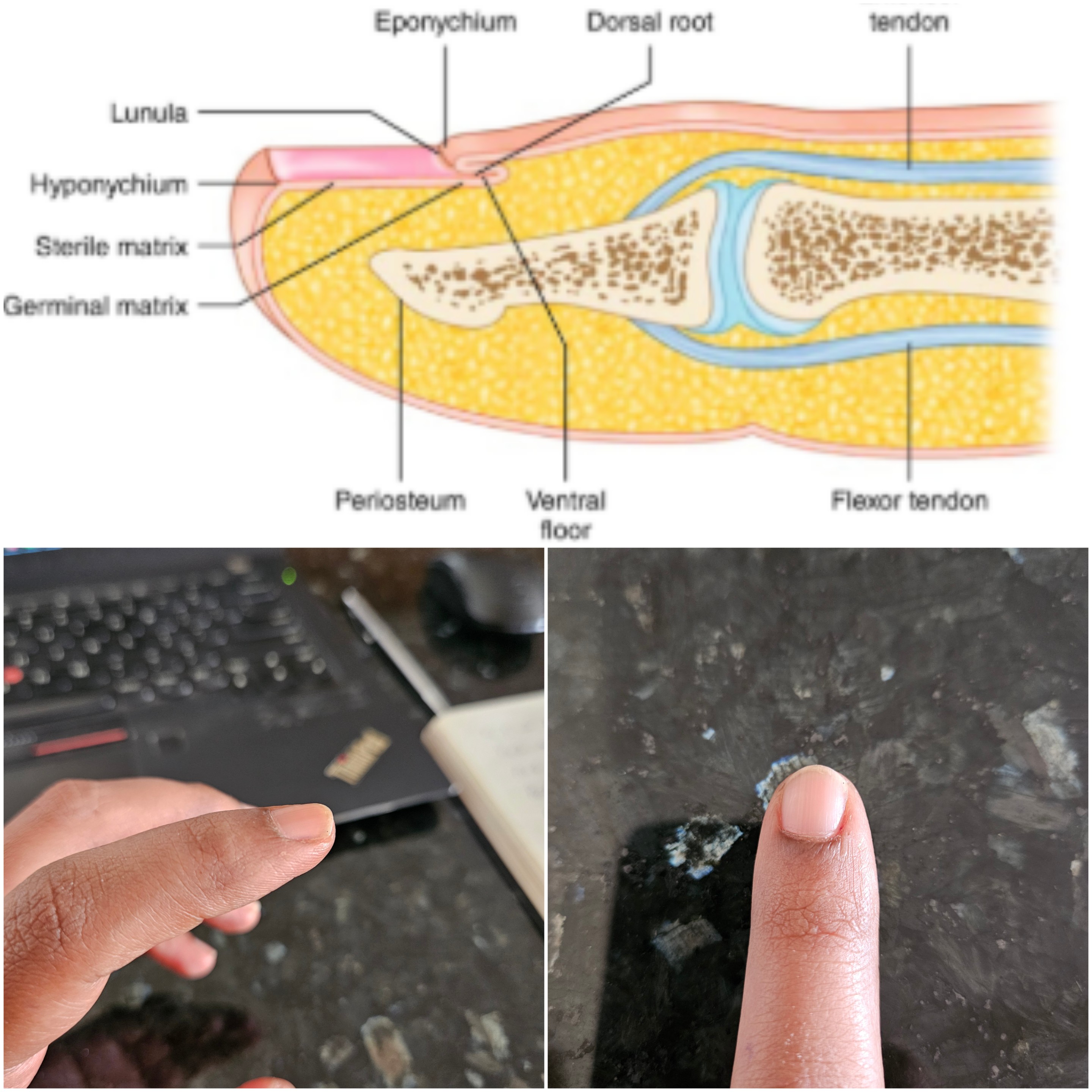
The fingertip is comprised of four primary structural pillars:
1. The Volar Pulp
The pulp is a specialized shock-absorbing cushion composed of subcutaneous fat arranged in tightly bound, vertical fibrous septae. These septae anchor the skin directly to the periosteum of the distal phalanx. This unique arrangement prevents the skin from shearing or slipping when we grip objects. The pulp is also packed with sensory nerve endings, giving us our exquisite sense of touch and two-point discrimination.
2. The Nail Unit (Perionychium)
The nail is not merely cosmetic; it is crucial for functional grip. It acts as a rigid backboard, providing counter-pressure against the volar pulp when grasping small objects. The perionychium includes:
● The Nail Plate: The hard keratinized structure.
● The Nail Bed: Consisting of the proximal germinal matrix (which produces 90% of the nail plate) and the distal sterile matrix (which anchors the nail plate as it grows forward).
● The Eponychium and Paronychium: The surrounding skin folds that protect and frame the nail.
3. The Distal Phalanx
The terminal bone of the finger provides the structural skeleton. It is wide at its base (articulating at the DIP joint) and tapers into a flat, spade-like distal tuft. The bone supports both the nail bed dorsally and the pulp volarly.
4. The Vascular and Nerve Supply
Blood is supplied by the digital arteries, which form a dense anastomotic arcade at the level of the distal phalanx. Innervation is provided by the digital nerves, which branch into numerous tiny terminal twigs to provide high-fidelity sensory feedback to the brain.
Section 3: Classification of Fingertip Injuries
Classifying fingertip injuries is essential for planning the appropriate surgical technique. In hand surgery, several classification systems are utilized, but two are highly critical when evaluating distal amputations and tissue loss: The Tamai Classification and the Allen Classification.
The Tamai Classification
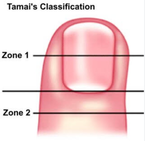
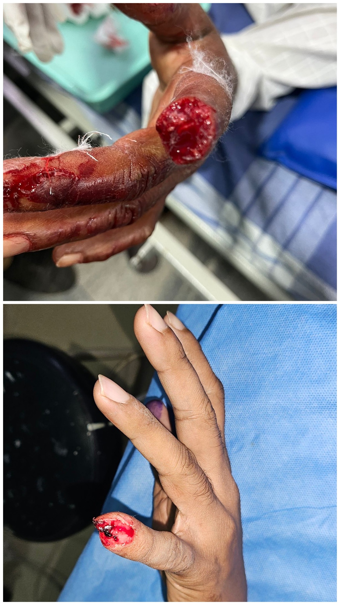
Developed by pioneering hand surgeon Sawaji Tamai, this system is specifically designed to classify amputations of the distal phalanx. It divides the terminal segment of the digit into two distinct zones:
● Tamai Zone I (Distal half): This zone extends from the mid-nail bed to the very tip of the finger. Injuries here involve only the distal pulp and the distal half of the nail bed. Because the bone loss is minimal or confined to the very tip of the distal tuft, these injuries are prime candidates for localized reconstructive flaps that preserve maximum length.
● Tamai Zone II (Proximal half): This zone extends from the distal interphalangeal (DIP) joint to the mid-nail bed. Injuries in Zone II represent a more severe class of trauma, involving the germinal matrix of the nail, the insertion of the extensor/flexor tendons, and a substantial portion of the distal phalanx.
At our Sarjapura Road center, Dr. Amrika Seshadri analyzes the exact boundaries of the Tamai zones to determine if a patient requires a local advancement flap, a cross-finger flap, or a conservative revision of the amputation site.
The Allen Classification
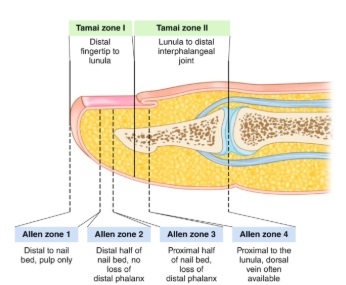
The Allen classification categorizes fingertip injuries based on the plane of injury and the anatomical structures involved:
● Type I: Involves only the pulp of the fingertip, sparing the nail bed and the distal phalanx.
● Type II: Involves the pulp and the nail bed, but spares the distal phalanx.
● Type III: Involves the pulp, the nail bed, and a partial loss of the distal phalanx (bone exposure).
● Type IV: Represents an amputation proximal to the lunula (the half-moon shape of the nail), involving the distal phalanx, nail matrix, and potentially the DIP joint.
Section 4: Clinical Evaluation and Initial Management
When a patient arrives at our Level 3A Superspeciality center with a fingertip injury, a structured, urgent clinical evaluation is initiated.
Step 1: Detailed Patient History
We document:
● Mechanism of Injury: Was it a clean cut (guillotine), a crush injury (door slam), or an industrial machine avulsion? Crush and avulsion injuries cause microvascular damage extending far beyond the visible wound edges, which dictates our choice of flap.
● Patient Profile: Age, hand dominance, occupation (e.g., manual laborer, office worker, professional musician), and medical history (smoking, diabetes, peripheral vascular disease greatly affect flap survival).
Step 2: Physical Examination
● Vascular Assessment: Assessing capillary refill, skin color, and active bleeding.
● Sensory Evaluation: Testing sensation wherever possible prior to administering local anesthetic.
● Structural Integrity: Checking for exposed bone, tendon involvement, or disruption of the nail matrix.
Step 3: Radiographic Assessment
X-rays of the affected hand (AP and lateral views) are mandatory to check for:
● Fractures of the distal phalanx.
● Dislocation of the DIP joint.
● Retained foreign bodies.
Step 4: First Aid and Stabilization
Before arriving at a specialized center, patients should follow these basic guidelines:
1. Stop the Bleeding: Apply direct, firm pressure with a clean cloth or sterile gauze. Do not use tight tourniquets or rubber bands at home, as this can starve the healthy tissues of oxygen.
2. Cleanse the Wound: Rinse gently with clean water or sterile saline. Do not scrub aggressively.
3. Elevate: Keep the hand elevated above the level of the heart to reduce swelling and throbbing.
4. Preserve Amputated Parts: If a portion of the finger has been cut off, wrap it in damp sterile gauze, place it inside a sealed plastic bag, and place that bag on a bed of ice. Do not let the amputated part touch ice directly, as freezing destroys the tissues. Bring the part to our clinic; even if replantation is not performed, the tissue can sometimes be used as a "spare part" skin graft.
Section 5: The Philosophy of Reconstructive Salvage vs. Replantation
Fingertip reconstruction is an exercise in balancing functional outcomes, aesthetic desires, and surgical risks. One of the most common questions patients ask is whether their severed fingertip can be reattached (replanted).
Why Dr. Amrika Seshadri Does Not Perform Replantation
Replantation is a highly specialized microvascular procedure where a surgeon attempts to sew back an amputated digit by anastomosing blood vessels that are often less than 1 millimeter in diameter. While it sounds ideal, replantation of fingertips (especially in Tamai Zone I) is highly controversial and often impractical for several reasons:
● Vascular Size: In the distal fingertip, the blood vessels are extremely small, tortuous, and prone to clotting (thrombosis).
● High Failure Rates: Replantation of crushed or bruised fingertips has a high failure rate, which can lead to prolonged hospitalization, repeat surgeries, and eventually, late amputation.
● Intense Post-operative Burden: Replantation requires a patient to remain hospitalized for several days with intensive monitoring, strict bed rest, and blood-thinning medications.
● Surgeon preference: Dr. Amrika has not trained extensively in Supermicrosurgery and therefore cannot, in good conscience, perform something that takes over a decade of persistent practice to execute.
Reconstructive Salvage: The Smart Alternative
Our philosophy at the Sarjapura Main Road clinic focuses on reconstructive salvage. By utilizing advanced local tissue transfers (flaps), we achieve outstanding, highly reliable results.
| Feature | Microvascular Replantation | Reconstructive Salvage (Flap Surgery) |
|---|---|---|
| Surgical Time | 4 to 8 hours | 45 minutes to 2 hours |
| Anesthesia | General or extensive regional block | Local or digital block |
| Hospital Stay | 5 to 7 days | Day-care procedure (discharge same day) |
| Reliability | Variable (highly susceptible to vessel clotting) | Extremely high (predictable blood supply) |
| Recovery Time | Months (with intensive hand therapy) | 3 to 6 weeks |
| Sensory Recovery | Often delayed or incomplete | Excellent (retains local nerve supply) |
| Key Advantage | Retains original skeletal length | Shorter recovery, less pain, rapid return to work |
Section 6: Advanced Reconstructive Techniques at Our Center
When a patient presents with a fingertip injury where bone is exposed or a simple skin graft is insufficient, we deploy specialized flap techniques. A flap is a unit of tissue that carries its own blood supply. Here are the cornerstone procedures performed by Dr. Amrika Seshadri:
1. The Atasoy Flap (Volar V-Y Advancement Flap)
The Atasoy flap (first described by Dr. Erdogan Atasoy in 1970) is an elegant, highly effective local flap used primarily for transverse or dorsal-oblique fingertip amputations with exposed bone (typically Tamai Zone I and Allen Type III injuries).
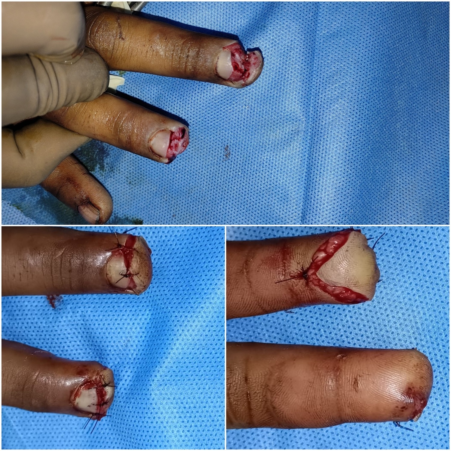
● Surgical Design: A triangular incision is made on the volar aspect of the fingertip. The base of the triangle is located at the distal wound margin, and the apex points proximally, extending toward the distal interphalangeal (DIP) joint crease.
● How it Works: The skin and subcutaneous tissue of the triangle are incised down to the periosteum of the distal phalanx. Crucially, the delicate fibrous septae anchoring the skin to the bone are gently released, while preserving the deep vascular connections from the digital artery branches. The triangular flap is then slid forward (advanced) over the exposed bone of the fingertip.
● The Closure: Once advanced, the skin edges are closed. The proximal stem of the wound is sutured in a straight line, transforming the original "V" incision into a "Y" configuration. This is why it is called a "V-Y" advancement flap.
● Advantages of the Atasoy Flap:
○ Like with Like: It uses the highly specialized, sensate skin of the volar pulp to cover the tip.
○ Maintains Length: It avoids the need to grind down or shorten the bone.
○ Sensory Preservation: It preserves the local nerves, meaning the fingertip retains almost normal feeling and touch.
○ Aesthetics: It creates a natural-looking, rounded fingertip contour.
2. The Cross-Finger Flap
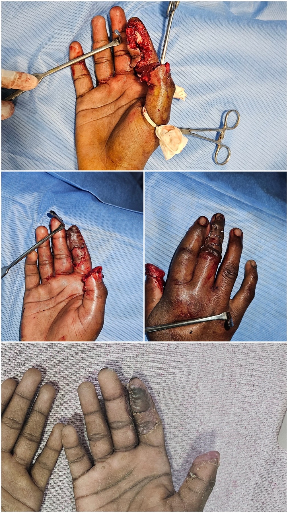
For larger, complex fingertip injuries—particularly volar-oblique amputations where a massive portion of the soft-tissue pulp is lost but the dorsal skin remains intact—the cross-finger flap is the gold standard.
● Surgical Design: This is a two-stage regional flap. It involves borrowing skin and subcutaneous tissue from the dorsal (back) side of an adjacent finger (usually the middle finger to repair the index, or the ring finger to repair the little finger) and transferring it like a "hinged door" to cover the wound on the injured finger.
● Stage 1 (The Attachment): The donor flap is raised from the back of the neighboring finger, leaving one side attached to maintain its blood supply. The flap is flipped over and sutured onto the raw fingertip wound of the injured finger. The fingers are temporarily bound together. To cover the raw dorsal area where the flap was taken, a small skin graft (usually harvested from the forearm or inner arm) is placed.
● Stage 2 (The Division): After approximately 2 to 3 weeks, the blood vessels from the injured finger grow into the flap, making it self-sufficient. At this stage, a brief outpatient procedure is performed to cut the connection between the two fingers, and the flap is neatly inset and tailored to form a new fingertip.
● Advantages of the Cross-Finger Flap:
○ Provides high-quality, durable skin coverage for extensive, deep volar defects.
○ Prevents joint contractures and scarring.
○ Provides a highly cushioned, stable pad for pinching.
Section 7: Rehabilitation, Scar Management, and Long-Term Care
A successful fingertip reconstruction does not end when the sutures are removed. At our Level 3A Superspeciality center on Sarjapura Main Road, we guide patients through a meticulous, multi-phase rehabilitation protocol to ensure optimal recovery.
1. Early Mobilization and Edema Control
● Elevation: During the first week, keeping the hand elevated above heart level is crucial to minimize throbbing and swelling.
● Adjacent Joint Movement: We encourage early movement of the proximal joints (the PIP, MCP, wrist, and elbow) to prevent stiffness in the rest of the hand.
2. Desensitization Therapy
Once the flap is fully healed (typically around week 3 or 4), patients often experience temporary hypersensitivity (hyperesthesia) or numbness. Desensitization is a crucial retraining process for the brain:
● Texture Stimulation: Gently rubbing the healed fingertip with different materials (such as soft cotton, silk, wool, and eventually rougher fabrics like denim) for 5-10 minutes, several times a day.
● Particle Immersion: Dipping the finger into bowls filled with rice, beans, or sand to normalize sensory feedback.
● Temperature Exposure: Alternating contact with warm and cool surfaces.
3. Scar Management
To prevent thick, painful scars or contractures:
● Silicon Gel Sheets: Applying medical-grade silicone sheets over the scar to soften it.
● Scar Massage: Massaging the healed surgical line in circular motions using a gentle moisturizer. This breaks up deep adhesions and prevents the skin from sticking to the bone underneath.
Section 8: Patient Testimonials and Case Insights
Case Study 1: Reconstructing a Mechanic's Index Finger
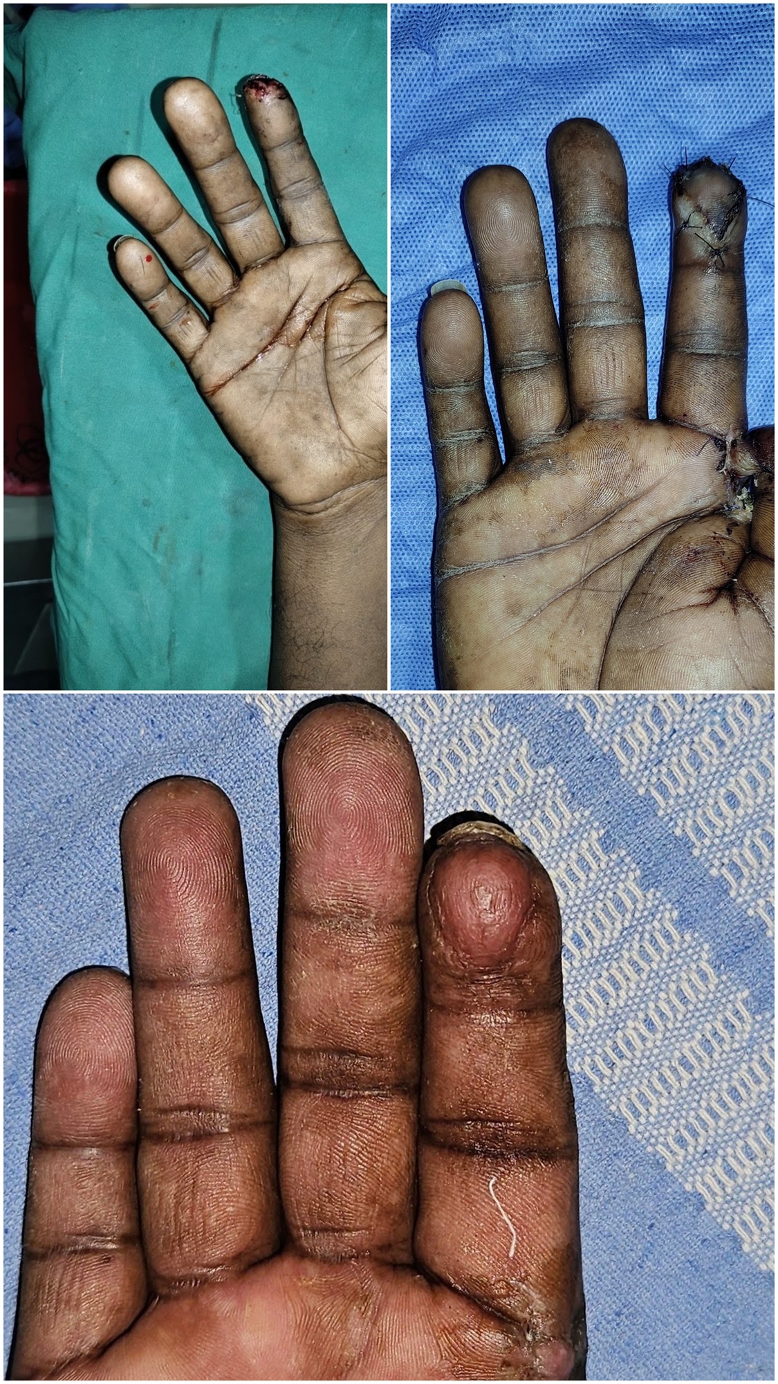
● Patient: Rajesh, a 34-year-old auto mechanic from Sarjapura.
● Injury: Sustained an Allen Type III, Tamai Zone I crush amputation of his left hand index finger in a hydraulic press. The bone was exposed, and there was significant loss of the volar pulp.
● Treatment: Dr. Amrika Seshadri performed an immediate Atasoy Volar V-Y Advancement Flap under local anesthesia at our clinic. The bone was preserved, the nail bed was stabilized, and the flap was advanced to cover the tip.
● Outcome: Within 4 weeks, Rajesh returned to light duties. By week 8, his two-point discrimination in the reconstructed finger was a remarkable 5mm (near-normal), allowing him to handle small nuts and bolts with ease.
Case Study 2: Saving the Ring Finger of a Software Engineer
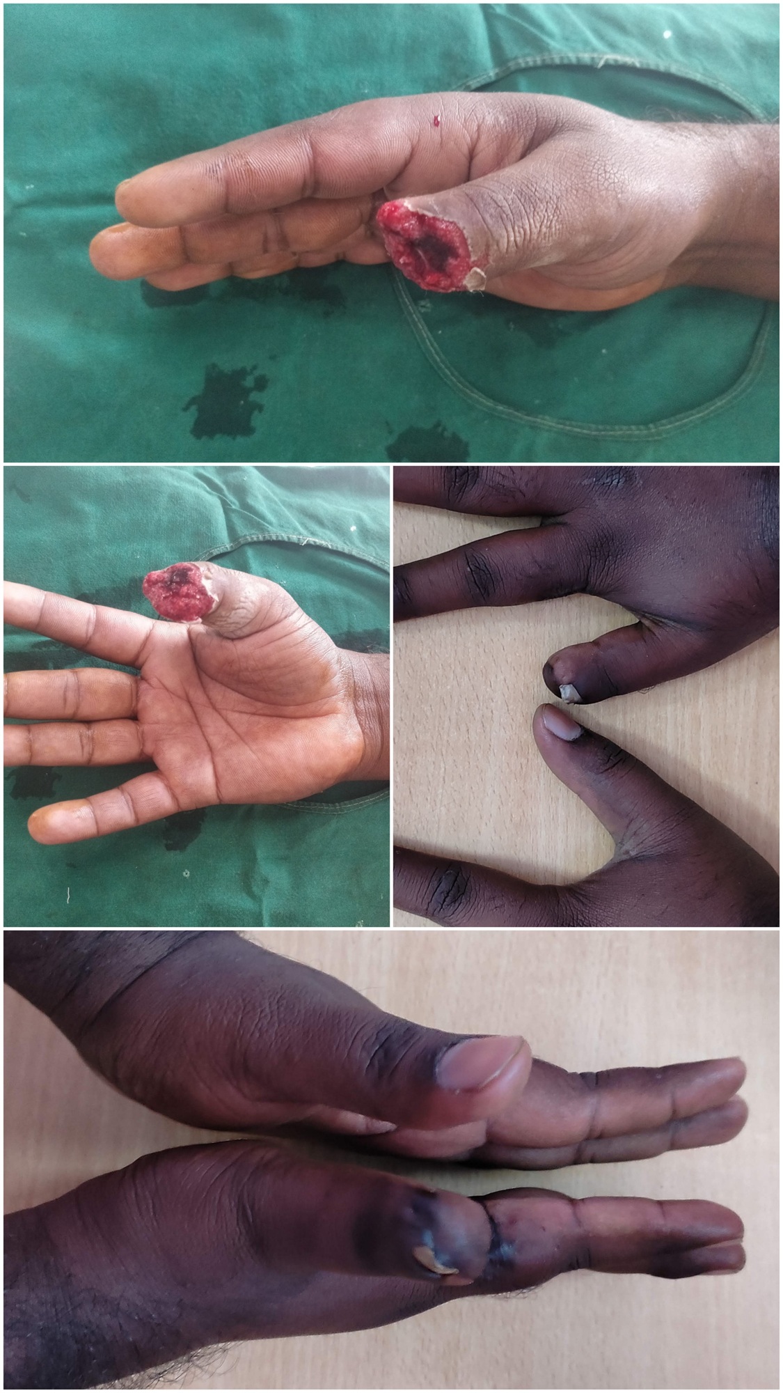
● Patient: Manjunath, a 28-year-old software developer.
● Injury: Severe bike chain accident resulting in a deep volar-oblique tissue loss of his right thumb. The bone was exposed, and there was no local skin left on the thumb to advance.
● Treatment: Dr. Amrika designed a Cross-Finger Flap borrowing tissue from the dorsum of his index finger. Stage 2 division was completed on day 21.
● Outcome: Manjunath achieved a highly aesthetic, well-padded fingertip that allows her to type seamlessly without any pain or cold sensitivity. Bonus- he developed a small nail from remnants of his germinal matrix, contributing to his aesthetic joy.
Section 9: Extensive Frequently Asked Questions (FAQ)
General FAQs
Q1: What is a "Level 3A Superspeciality" center, and why is it important for fingertip injuries?
A Level 3A Superspeciality center is an advanced medical facility equipped with specialized surgical suites, high-definition magnification systems, and dedicated hand rehabilitation services. For fingertip injuries, this means we have the precise instruments and specialized materials (such as ultra-fine sutures and specialized dressings) required to work on delicate structures like the nail matrix, nerves, and small blood vessels.
Q2: How long does a fingertip reconstruction procedure take?
Depending on the complexity, a local flap (like an Atasoy flap) takes between 45 to 75 minutes. A regional flap (like the first stage of a cross-finger flap) may take 1.5 to 2 hours. These are typically performed as day-care procedures, meaning you can go home the same day.
Q3: What type of anesthesia is used for these procedures?
Most fingertip reconstructions are performed under a digital nerve block (local anesthesia administered at the base of the finger). This completely numbs the finger while keeping you fully awake, comfortable, and avoiding the risks and recovery time of general anesthesia. For highly anxious patients or children, light sedation can be provided.
Q4: Why does Dr. Amrika Seshadri not perform replantations?
Replantation of fingertips (sewing back an amputated tip) is a highly intensive procedure with a high failure rate in crushed or bruised tissues, and it carries a heavy post-operative burden. Dr. Amrika's 11 years of clinical experience have shown that reconstructive salvage using advanced local flaps (such as Atasoy or cross-finger flaps) delivers highly reliable, durable, functional, and pain-free fingertips with significantly less downtime, lower costs, and almost no risk of total tissue failure.
Q5: Will my nail grow back normally after a fingertip injury?
If the injury is in Tamai Zone I and the germinal matrix (located under the cuticle) is undamaged, the nail will grow back, though it may take 3 to 6 months to grow completely. If the nail bed was lacerated and carefully repaired, the nail is likely to grow back smoothly. However, if there is severe damage to the germinal or sterile matrix, some minor nail deformities (such as ridges or a split nail) may occur.
Q6: How long will I be off work after surgery?
● Office/Desk Jobs: You can often return to work within 3 to 5 days, provided your hand is kept protected and elevated.
● Manual Labor/Heavy Gripping: You may need 4 to 6 weeks to allow the flap to mature and gain full mechanical strength before resuming heavy lifting or wet work.
Q7: Who is the best surgeon for fingertip amputation injuries in Bengaluru?
This question is best answered in 2 parts:
1. The most qualified surgeon for this procedure is a Plastic Surgeon. This is because a Plastic Surgeon is obsessed with minimal scarring and smooth recovery. Dr. Amrika Seshadri has been engaged in providing this service for the past 11 years and has a great deal of experience.
2. Patient comfort and confidence is paramount for this procedure to work perfectly. Choose a qualified Plastic Surgeon who helps you understand your best solution.
Q8: Can this surgery be covered under Insurance? Since the Surgeon is a Plastic Surgery, won’t this be considered Cosmetic Surgery?
Fingertip injuries, though minor in size, can have manifold QOL issues. Therefore, nailbed repairs, cross finger flaps and locoregional fingertip flaps are considered Reconstructive in nature and Medically Essential. This has complete coverage under all major Insurances. At Dr. Amrika’s Aesthetics, we provide end-to-end Insurance reimbursement services to every patient who chooses us for their treatment.
Medical & Technical FAQs
Q7: What is the primary difference between Tamai Zone I and Zone II injuries?
Tamai Zone I injuries occur on the distal half of the phalanx (from mid-nail bed to the tip) and are easily amenable to local flaps like the Atasoy V-Y flap. Tamai Zone II injuries occur proximally (from the DIP joint to the mid-nail bed) and involve the insertion of the tendons and the nail matrix. Zone II injuries often require more complex coverage, such as cross-finger flaps, or revision amputation if the joint is unsalvageable.
Q8: How does an Atasoy flap preserve sensation?
The Atasoy flap is advanced along with its deep subcutaneous tissue, which contains the terminal branches of the digital nerves and arteries. Because the nerves are not cut but merely mobilized along with the skin, the flap retains its original sensory pathways, allowing for excellent recovery of feeling.
Q9: What are the risks of a V-Y advancement flap?
While highly reliable, potential risks include:
● Partial Flap Necrosis: If the blood supply is compromised (more common in heavy smokers or diabetics).
● Hook Nail Deformity: If the flap is not advanced far enough to support the nail bed, the nail may curve downward over the tip of the finger. Dr. Amrika uses meticulous anchoring techniques to prevent this.
● Hypersensitivity or Numbness: Usually temporary and manageable with desensitization therapy.
Q10: Does a cross-finger flap leave the donor finger damaged?
The donor site on the adjacent finger is covered with a thin skin graft. While it will have a slightly different color and texture than the original skin, it heals completely and does not affect the movement, strength, or function of the donor finger.
Q11: Can a fingertip injury heal without surgery?
Very minor injuries (Allen Type I, involving only small skin loss without bone exposure) can heal beautifully on their own through a process called healing by secondary intention. This involves regular cleaning and specialized dressings. However, if bone is exposed (Allen Type III & IV), leaving the wound open carries a high risk of bone infection (osteomyelitis) and chronic pain, making surgical flap coverage essential.
Q12: How do I manage chronic cold sensitivity after a fingertip injury?
Cold intolerance is a common side effect of hand injuries, occurring because of hypersensitive nerve endings. It usually improves gradually over 1 to 2 years. Keeping your hands warm with gloves during cold weather, avoiding sudden temperature changes, and continuing desensitization exercises can help manage this symptom.
Section 10: Conclusion and Next Steps
Sustaining a fingertip injury can be a highly stressful and painful experience. However, with the right surgical expertise and timely intervention, these injuries can be managed with outstanding outcomes.
If you or a loved one has sustained a fingertip injury, remember that preserving length, restoring touch, and avoiding chronic pain depend heavily on the skill of the treating surgeon. With 11 years of dedicated practice in plastic and reconstructive surgery, Dr. Amrika Seshadri provides world-class care right on Sarjapura Main Road.